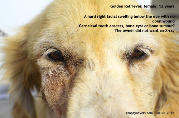
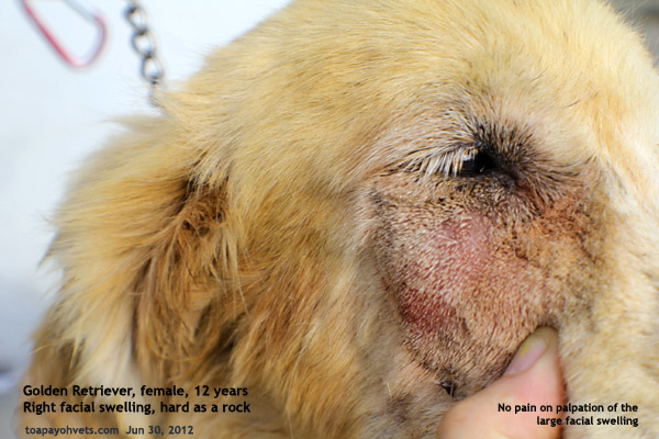
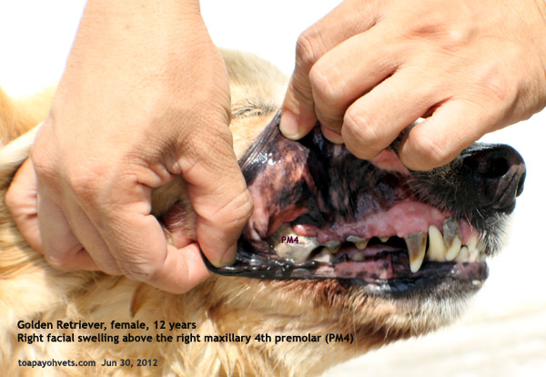
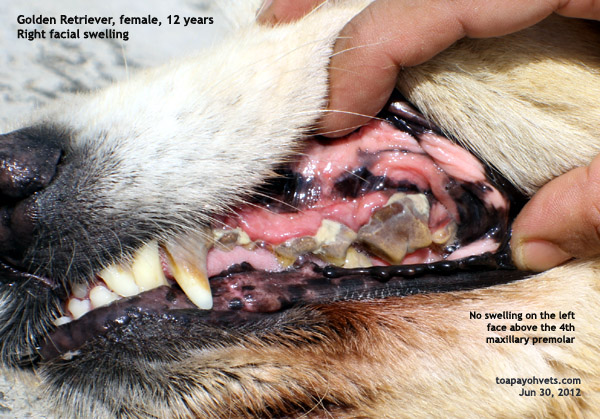
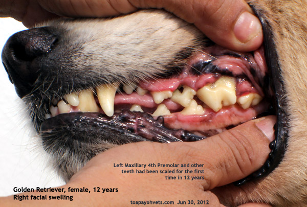
The 12-year-old Golden Retriever had a big hard lump on the right face, below the eye, in the position of a carnaissal tooth abscess fistula. At first sight, I diagnosed a carnaissal tooth abscess as this is the most common location and problem in older dogs that do not have any dental work done in 12 years! Dr Daniel said it could be a bone cyst or tumour.
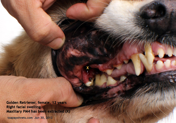
As the owner did not want any X-ray to be done, Dr Daniel extracted the right maxillary 4th premolar.
I noted that the roots are shrivelled and blackened but I was not present during the dental work. According to Dr Daniel, this would not be a carnaissal tooth abscess. "I have seen two cases of bone cysts in Australia" he was doing internship then.
"Did you see the bone cyst in a similar location, below the eye?" I clarified with him later.
"Not
in this location but bone cyst can occur in anywhere as a hard
swelling from the bone. Bone cyst is a differential
diagnosis."
"I have seen none in my past 30 years of practice," I said.
99% of my cases are small breeds as over 80% of Singaporeans
live in apartments and so Golden Retrievers are uncommon
patients and so I have not seen one with carnaissal tooth
abscess in this breed.
 |
 |
 |
 |
 |
 |
 |
 |
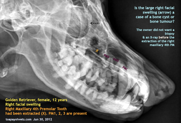
So, is this a case of a carnaissal tooth
abscess or not? The owner did not want histopathology or
biopsy but agreed to X-rays after the tooth extraction. I have
cropped the X-rays to focus on the relevant areas and to
scrutinise them. What do you think?
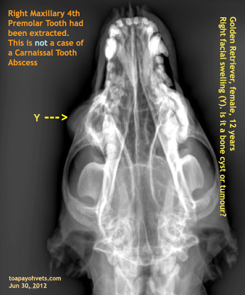
X-ray after extraction of the maxillary PM4 showed a large
dense globular lump (Y). It is hard to say whether it is a
bone cyst, bone tumour or encapsulated abscess of the root of
the carnaissal tooth.
MY HYPOTHESIS
Due to its unique location, I would say this hard lump is
associated with the carnaissal tooth infection going on for
many years. This dog did not have dental work for the last 12
years and the tooth root had rotted away sending bacteria into
this area, infecting the bone. The reaction is a hard lump
which the owner noticed recently. The dog was still "eating"
and the owner consulted us for the lump.